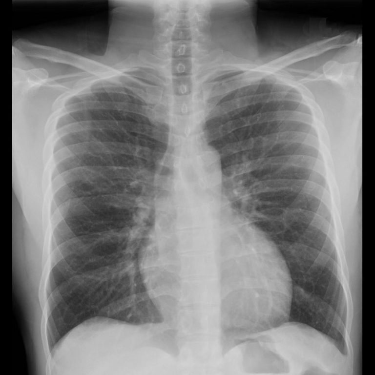

However, a pneumothorax is more clearly seen in an expiratory film rather than in the inspiratory film. In future studies, an association between malpositioning and adverse events should be investigated.Interpretation of the film taken in expiration is difficult because the lung bases appear hazy and the heart looks enlarged. This is relevant because malpositioned devices may be related to adverse events. Malpositioning of central venous catheters and endotracheal tubes is frequently identified in radiograms of patients in an intensive care unit. Of the endotracheal tubes, 19.9% were malpositioned, while all the tracheostomy tubes were correctly positioned. Of the central venous catheters that were identified, 33.6% of the subclavian and 23.8% of the jugular were malpositioned. Several medical devices were identified, including monitoring leads, endotracheal and tracheostomy tubes, central venous catheters, pacemakers and prosthetic cardiac valves. Out of the 2,312 thoracic radiograms analyzed, 568 were included in this study.

The radiograms were evaluated by an independent observer. One radiogram per admission was selected for analysis. All admissions in which at least one thoracic radiogram was performed in the intensive care unit and in which at least one medical device was identifiable in the thoracic radiogram were included. All the thoracic radiograms performed in the intensive care unit of our center over an 18-month period were analyzed. To identify and evaluate the correct positioning of the most commonly used medical devices as visualized in thoracic radiograms of patients in the intensive care unit of our center.Ī literature search was conducted for the criteria used to evaluate the correct positioning of medical devices on thoracic radiograms.

The goal of this review is to summarize the data regarding the use of leadless cardiovascular devices including the CardioMEMS device, implantable loop recorder, and RV leadless pacemaker, and to present cases demonstrating their utility and normal imaging features. As these devices become more widely used, it is important for radiologists to become familiar with the normal imaging features and potential complications. There is limited published data regarding the imaging features of these devices. Additionally, hemodynamic monitoring devices have shown utility in monitoring patients with aortic aneurysms after endovascular aortic repair (EVAR) for early detection of Type I and Type II endoleaks. Cardiovascular devices including the CardioMicroelectromechanical (CardioMEMS) device, implantable loop recorder, and right ventricular (RV) leadless pacemaker are now widely used for treatment and monitoring of advanced cardiac conditions, as many of these devices have been shown to significantly improve patient outcomes. The obtained results allowed for the continuation of the pilot study for augmented research and material properties analysis.Ĭardiovascular devices and hemodynamic monitoring systems continue to evolve with the goal of allowing for rapid clinical intervention and management. A significant increase in quantity was achieved using laser power with a minimum power of 15 mW. The relationship between the laser power and the characteristics of the particles remaining in the laser irradiation area has been established. Attempts to reduce deposited gold coating to the size of Au nanoparticles (Au NPs) and to fuse them into the groove using a laser beam have been successfully completed. The visibility of the constituted coating was checked by a commercial grade X-ray that is commonly used in hospitals.

The evaluation of gold particle size reduction was performed with scanning electron microscopy (SEM) (secondary electron (SE) and quadrant backscatter electron detector (QBSD) and energy dispersive spectroscopy (EDS) analysis. The initial material analysis was conducted by power–depth dependence using confocal microscopy. The process was suited to avoid intensive surface modification that could compromise the mechanical properties of fragile cardiovascular devices. The energy of the laser beam stating the presence of polymer bond thermalisation with remelting due to high temperature was also taken into the account. The structures were investigated by employing surface topography in the presence of micron and sub-micron features. Biomimetic pattern and parameters optimization were key properties of the design for biomedical application. To fuse particles into polymer, the raw surface of PEEK was sputtered with 99.99% Au and micromachined by an A-355 laser device for gold particle size reduction. This paper analyses the possibility of obtaining surface-infused nano gold particles with the polyether ether ketone (PEEK) using picosecond laser treatment.


 0 kommentar(er)
